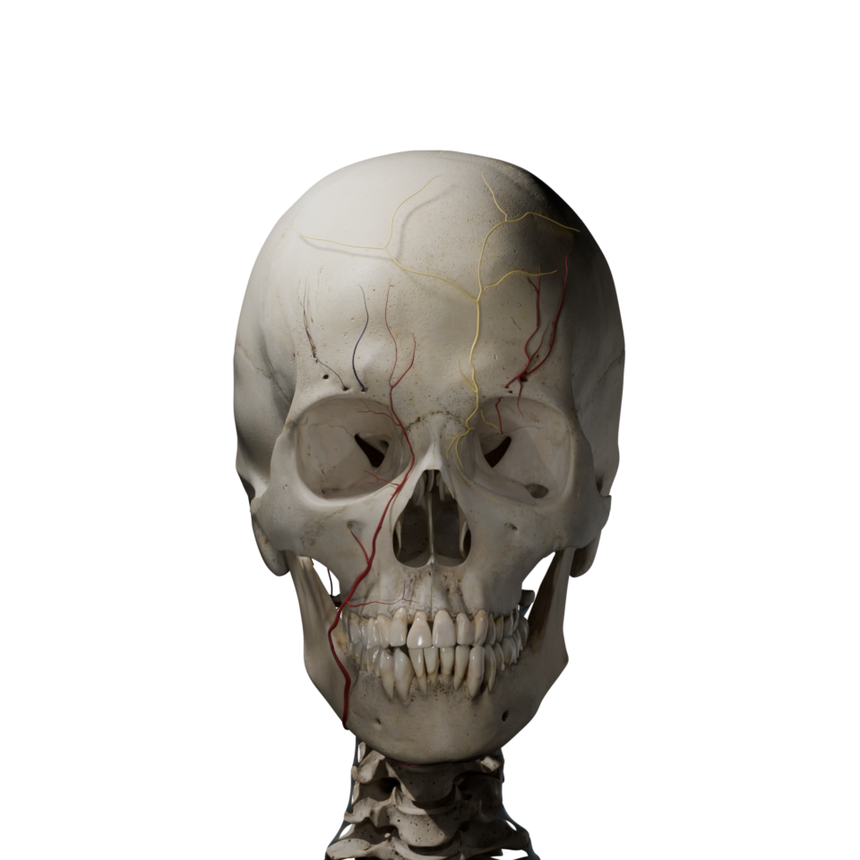
Hyperrealisticillustrations
Explore anatomy through detailed, medically accurate visuals that reveal depth, landmarks, and key structures, making complex anatomy easier to see, understand, and remember.

Explore anatomy through detailed, medically accurate visuals that reveal depth, landmarks, and key structures, making complex anatomy easier to see, understand, and remember.
Practice with image-based questions that train recognition, strengthen understanding, and help anatomy concepts stay in memory for exams.
Start studying complex anatomical structures in the language of your choice.
The ulnar shaft is triangular and tapers distally. It has three borders and three surfaces.
The interosseous border faces the radius and provides attachment for the interosseous membrane, which links the forearm bones and transmits load. The posterior border is subcutaneous and palpable, and serves as an attachment for fascia and muscles such as flexor carpi ulnaris and extensor carpi ulnaris.
The anterior border is rounded and provides origin for deep flexors, including flexor digitorum profundus, and distally for pronator quadratus. The posterior surface provides attachment for posterior compartment muscles, including abductor pollicis longus, extensor pollicis longus, and extensor indicis distally.
A nutrient foramen is usually present on the anterior surface, transmitting vessels to the medullary cavity.
Next: 54.4 Distal end of the Ulna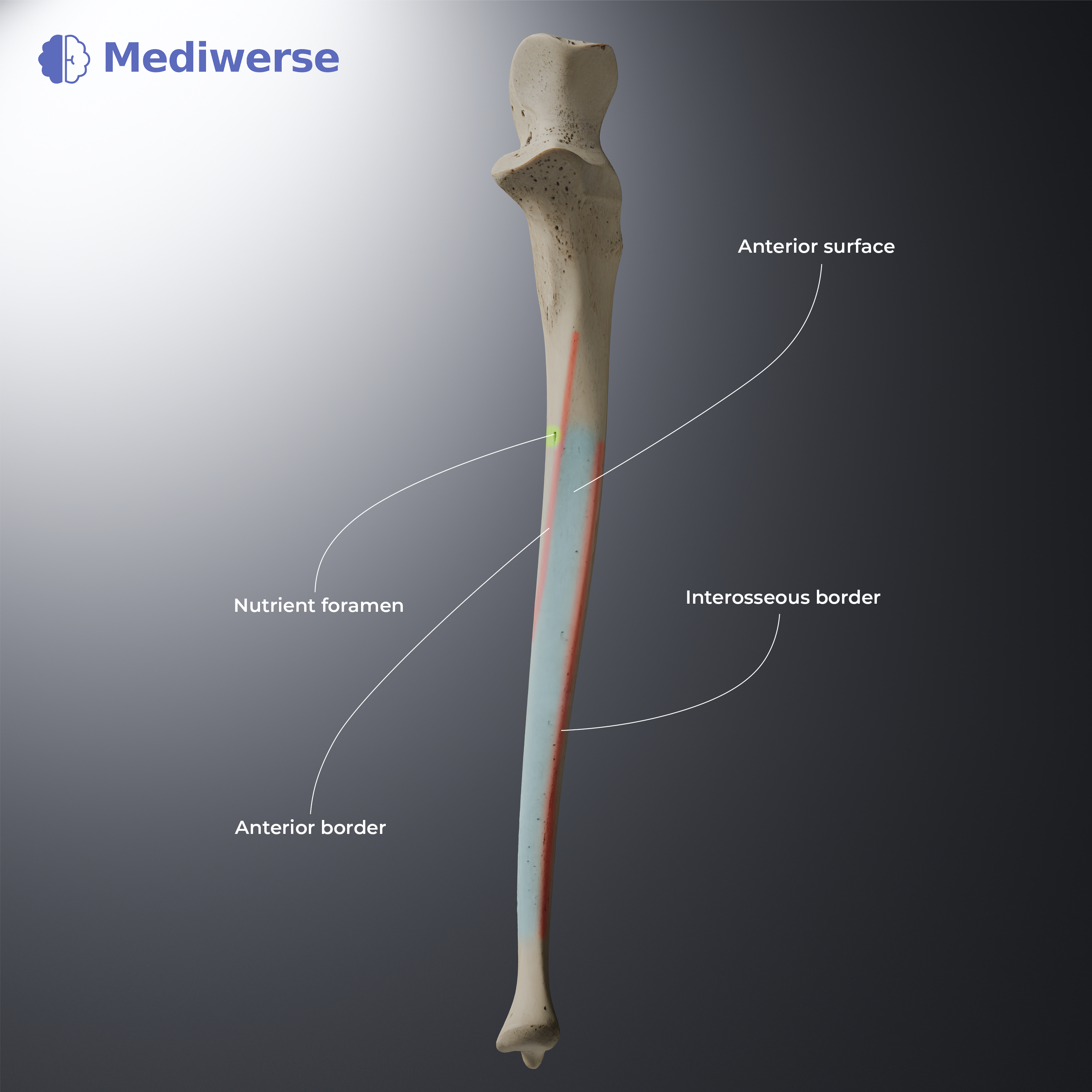
Access Mediwerse anywhere, anytime. It works just as well on every screen.
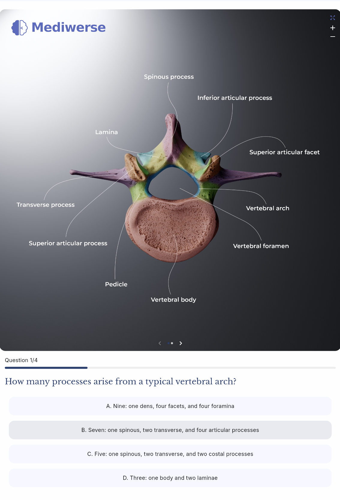
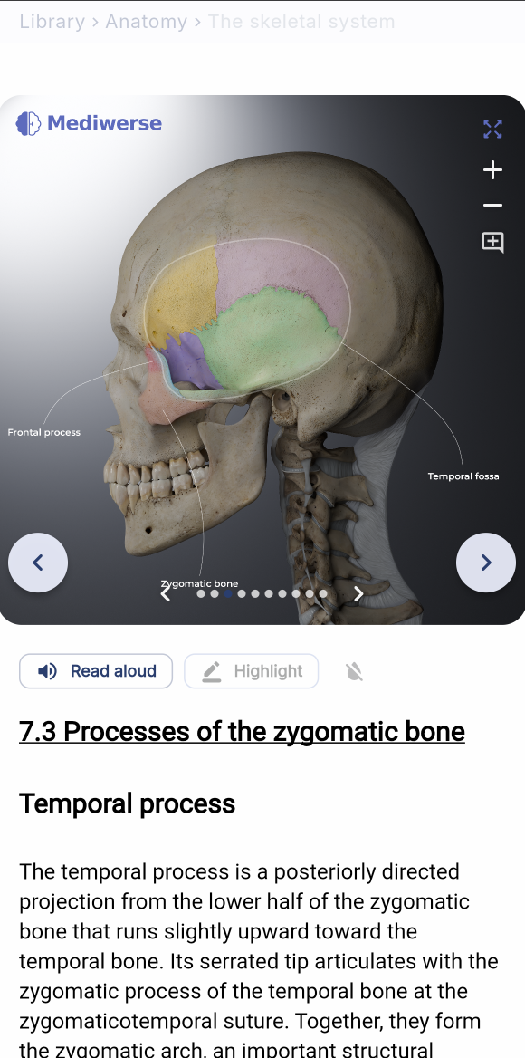
Create your own chapters with Mediwerse visuals. Write your own study notes, quizzes and share them with your friends.
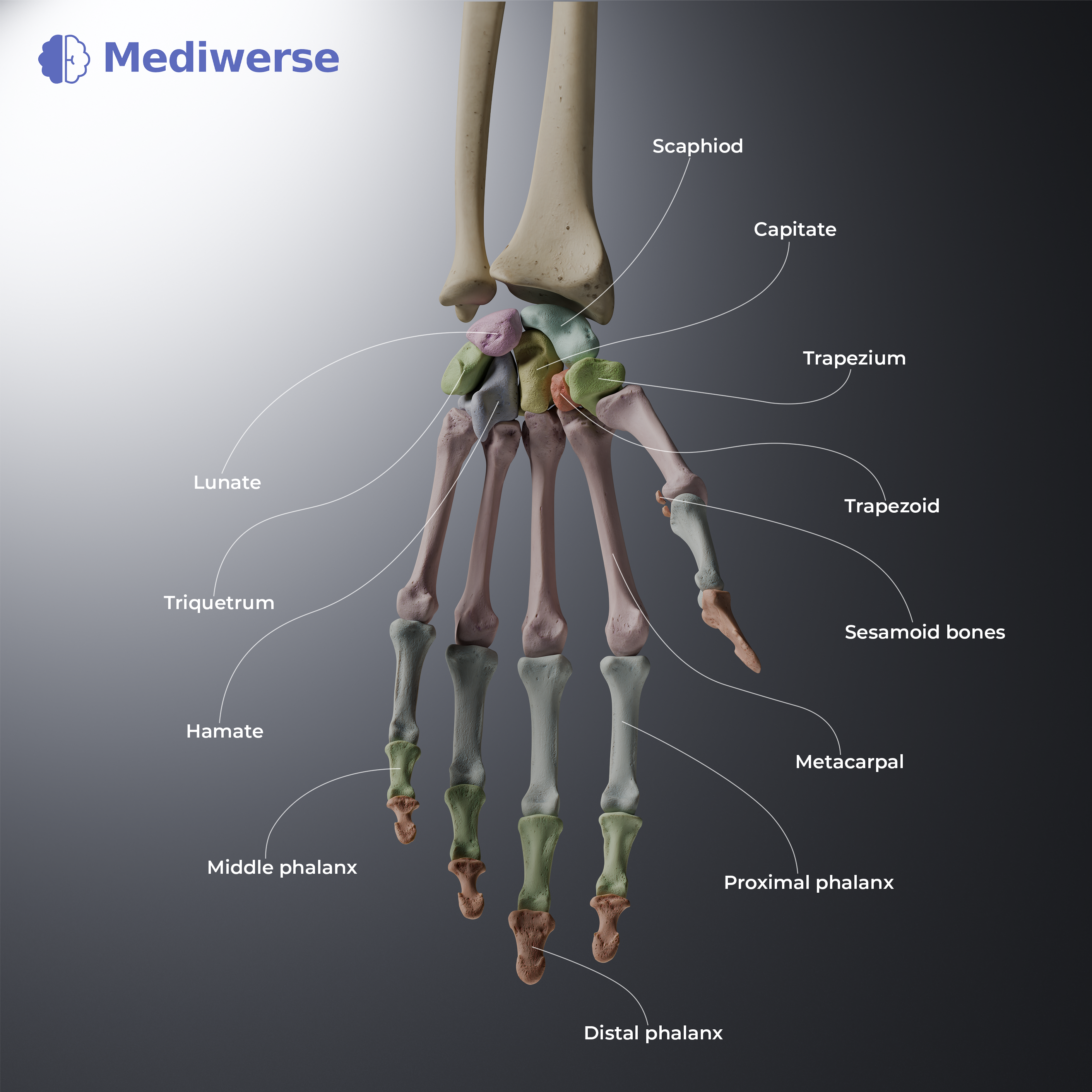
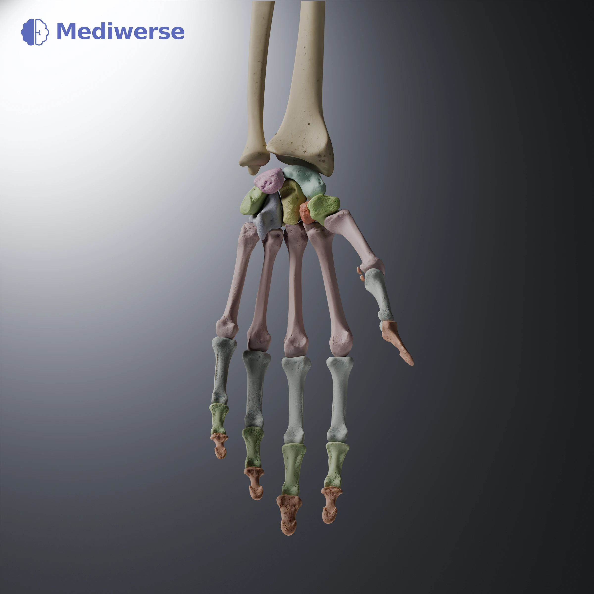
- Scaphoid: A boat-shaped bone on the thumb side; it is the most frequently fractured carpal bone.
- Lunate: A crescent moon-shaped bone sitting in the center.
- Triquetrum: A pyramidal bone on the pinky side of the wrist.
- Pisiform: A small, pea-shaped bone that rests on top of the triquetrum.
- Trapezium: A four-sided, saddle-shaped bone that forms the base of the thumb.
- Trapezoid: A smaller, wedge-shaped bone next to the trapezium.
- Capitate: The largest carpal bone, located right in the center.
- Hamate: A wedge-shaped bone on the pinky side characterized by a distinct bony hook.
See the anatomy clearly while you study it.
Study anatomy in your native language.
Create personalized tests for the exact topics you want to practise.
Review previously missed questions to strengthen weak areas and improve long term retention.
Create your own personalized study notes and quizzes using our hyperrealistic illustrations.
Share your own study notes and quizzes with your friends
Listen to the text while reviewing the illustrations.
Highlight important sections in the text so they are easier to find and return to.
Mediwerse brings visual anatomy learning, active recall, personal study notes and multilingual study tools into one focused platform for medical and healthcare students.
Mediwerse is an educational anatomy platform. It does not provide clinical diagnosis, treatment, or patient-specific medical advice.
Start FREE trial now